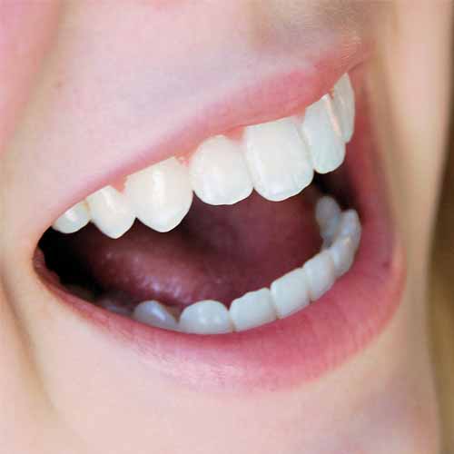
Dental radiographs
(X-rays) are essential, preventative, diagnostic tools that provide
valuable information not visible during a regular dental exam. Dentists
and dental hygienists use this information to safely and accurately
detect hidden dental abnormalities and complete an accurate treatment
plan. Without X-rays, problem areas may go undetected.
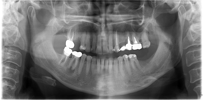
Dental X-rays may reveal:
- Abscesses or cysts.
- Bone loss.
- Cancerous and non-cancerous tumors.
- Decay between the teeth.
- Developmental abnormalities.
- Poor tooth and root positions.
- Problems inside a tooth or below the gum line.
Detecting and treating dental problems at an early stage can save you time, money, unnecessary discomfort, and your teeth!
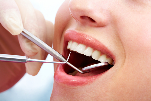
Are dental X-rays safe?
We
are all exposed to natural radiation in our environment. The amount of
radiation exposure from a full mouth series of X-rays is equal to the
amount a person receives in a single day from natural sources.
Dental
X-rays produce a low level of radiation and are considered safe.
Dentists take necessary precautions to limit the patient’s exposure to
radiation when taking dental X-rays. These precautions include using
lead apron shields to protect the body and using modern, fast film that
cuts down the exposure time of each X-ray.
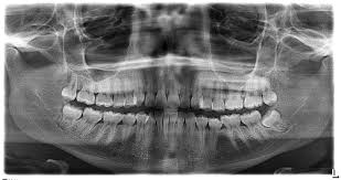
How often should dental X-rays be taken?
The need for dental X-rays depends on each patient’s individual
dental health needs. Your dentist and dental hygienist will recommend
necessary x-rays based on the review of your medical and dental history,
dental exam, signs and symptoms, age consideration, and risk for
disease.
A full mouth series of dental X-rays is recommended for new patients. A full series is usually good for three to five years. Bite-wing X-rays
(X-rays of top and bottom teeth biting together) are taken at recall
(check-up) visits and are recommended once or twice a year to detect new
dental problems.